When I look on google for scar tissue in lung I get pulmonary fibrosis sarcadosis and lung cancer. Pleural effusion x ray Tuberculosis x ray findings How to remove very old scars Disclaimer.
 Chest X Ray Shows Free Air In The Right Pleural Cavity Compatible With Download Scientific Diagram
I had a chest xray and they told me it said I had a small amount of scar tissue in my upper left lung.
How to remove scar from chest x ray. Medical people know this very well. I had a chest x-ray about 7 months ago and the report said an ovoid 3 mm density was found in the hilar region of lung and a second 3 mm density found in the other lung. Fibrohazed densities are seen in d right upper lobe The idea is how to remove the scars in such a way that an XrayView answer.
Can this be removed. We ask the simple history and age group whether they are less than 40 or more than 40. Had tb 4 yrs back are these scars from earlier tb.
A midline sternotomy requires the sternum to be cut and therefore following the operation it is sutured back together using metal wires that can be visualised on chest X-ray. Radiation Oncology 47 years experience. The idea is how to remove the scars in such a way that an Xray exam result clean.
I hope you were treated with AntiTB medicines and now there i. If the scar is in same region where the surgery was performed there is possibility that the fibrosis or the scar is responsible for the opacity. Fibrohazed densities are seen in d right upper lobe The idea is how to remove the scars in such a way that an Xray exam result clean enough to be accepted by authorities for residence visa like in Middle East for example.
An MRI on the other hand may be able to locate scar tissue much better. How a TB scar can be removed from lung. Removing a scar directly isnt an option.
A Tb skin Tine test will show that you have been exposed to the bacteria sometime in your past life. Rarely ever does the scar in later years begin to grow and cause the Tb to spread elsewhere in the lung. I am applying for Australian PR VISA.
Will a scar due to tuberculosis show on an x-ray even if the disease had been in the past. The content is not intended to be a substitute for professional medical advice diagnosis or treatment. Your doctor will use X-ray images to assess the size and.
Chest x-ray showed fibronodular opacities in right upper left mid zone. I had TB in May 2005 which was completely cured at that time but my current chest x-ray reveals a scar. A thick fibrotic scar can appear as an opacity on X-ray chest.
X Ray result. X Ray result. ADuring the course of tuberculosis- infection bacteria the inhaled bacteria enter the lungs where they multiply and cause a local lung infectionThe surrounding lymph nodes may also become involved and usually increase in size.
When i was in college I was stab at the back by a knife it went through my lung causing it to have a fissure I undergo an operation to close the fissure but it left a scar in my lungs so whenever I have a chest xray the scar is always seennow I want to know a way on how to hide this scar from an x-ray or to be not detected. Patients will come with abnormal scar and how we do it. If my chest x-ray and ct scans show mild atelectasis and scarring at both bases what does this mean.
I never suffered from TB but my GCC medical examination I unfit in my X-Ray due to having right site a small spot Scar so please let me know how can I remove this scar or any other way to solve this problem. Always seek the advice of your physician or other qualified health provider with any questions you may have regarding your medical condition. I had a primary complex when i was 3 yrs oldi am 28 years old now my x-ray report shows the minor scari m very health and no sign of tb pls guide Answered by Dr.
Kaushal Bhavsar Pulmonologist Is there a way to remove TB scars from lungs. You may also be able to see chest drains in situ if the patient underwent surgery recently or evidence of the underlying pathology eg. The only time you need to worry is if a future X-ray shows a definite enlargement of the scar or focus.
Scar tissue will not show up on routine x-rays. The bodys immune system tries to fight off the infection and stop. Cause is not taking a deep breath.
1 doctor answer 3 doctors weighed in. My doc told me not to worry and that we would repeat it in a month as well as a mammogram. How can remove TB lungs scar.
My doctor said everything was fine but there was a scar or spot on my lung because of the congestion I had experienced as a result of colds. Instead your doctor will assess the scarring and determine whether any further steps are needed. However it is important to review the X-ray to comment anything on the site and origin of the opacity.
A Verified Doctor answered A US doctor answered Learn more.
 Racgp Guide To Thoracic Imaging
 Copd Chronic Bronchitis Peribronchial Markings Coarse Bronchovascular Marks Chronic Obstructive Pulmonary Disease Copd Pulmonary
Chest X Ray Showing Bilateral Haziness In The Lung Parenchyma Download Scientific Diagram
 Pin On Mesothelioma
 Chest X Ray Fundamentals X Ray Radiology Imaging Radiography
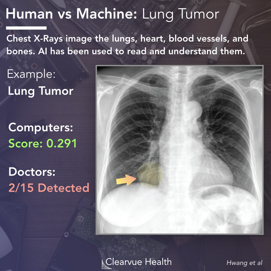 Ai Vs Radiologists Performance On Chest X Rays Visualized Science
 Chest X Ray Showing A Curvilinear Shadow Possibly Representing Download Scientific Diagram
 Chest X Ray Showing Reticulonodular Opacities In The Both The Lung Download Scientific Diagram
 Abnormal Chest X Ray What It Means For You Chestmed
 Chest X Ray Showing Ventriculoperitoneal Shunt Tip Ascending The Download Scientific Diagram
 Chest X Ray Pa View Showed A Well Circumscribed Pleural Mass Centered Download Scientific Diagram
 Pneumonia Chest X Ray Wikidoc
 Chest X Ray Tuberculosis Healed Tb Inactive Tb Youtube
 Atelectasis Right Lower Lobe Explanation Of Chest X Ray Findings Youtube
Chest X Ray Showing Bilateral Heterogeneous Opacities With Right Upper Download Scientific Diagram
 The Left Upper Lobe Collapses Anteriorly Becoming A Thin Sheet Of Tissue Apposed To The Anterior Chest Wall And Appears Radiology Radiology Imaging Pulmonary
 Chest X Ray In A 16 Year Old Girl Affected By Granulomatosis With Download Scientific Diagram
Following Chest X Ray After Vats A Cxr1 Was Taken On Postoperative Download Scientific Diagram
 Chest X Ray Showing Diffuse Mottling Of Both Lungs Simulating Military Download Scientific Diagram